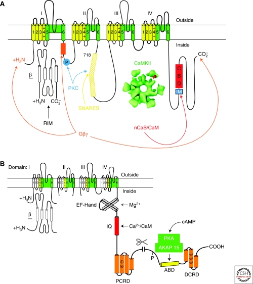
Figure 4.
Ca2+ channel signaling complexes. (A) The presynaptic Ca2+ channel signaling complex. A presynaptic Ca2+ channel α1 subunit is illustrated as a transmembrane folding diagram as in Figure 2. Sites of interaction of SNARE proteins (the synprint site), Gβγ subunits, protein kinase C (PKC), CaMKII, and CaM and CaS proteins are illustrated. IM, IQ-like motif; CBD, CaM binding domain. (B) The cardiac Ca2+ channel signaling complex. The carboxy-terminal domain of the cardiac Ca2+ channels is shown in expanded presentation to illustrate the regulatory interactions clearly. ABD, AKAP15 binding domain; DCRD, distal carboxy-terminal regulatory domain; PCRD, proximal carboxy-terminal regulatory domain; scissors, site of proteolytic processing. The DCRD binds to the PCRD through a modified leucine zipper interaction.

