Figure 2.
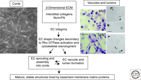
Fundamental contribution of the ECM-integrin-cytoskeletal signaling axis to the assembly of EC cords and lumens during vasculogenesis and angiogenesis. In this schematic, two models of EC morphogenesis are illustrated. In the left panel, confluent human microvascular EC monolayers were overlaid with a type I collagen gel and allowed to undergo cord formation, illustrating how collagenous ECM markedly converts an EC monolayer into an interconnecting network of cords. This process is dependent on the α1β1 and α2β1 integrins and the small GTPase, RhoA. In the right panels, human ECs are suspended as individual cells in 3-D collagen matrices and are fixed, stained, and photographed at 8 and 48 hr of culture, illustrating intracellular vacuoles and early lumen formation at 8 hr and interconnecting networks of EC-lined tubes at 48 hr. Bar equals 50 µm. The three panels on the far right side are cross-sections from plastic-embedded collagen gels revealing EC intracellular vacuoles (upper two right panels) or EC lumens (lower right panel). Bar equals 25 µm. The vacuole and lumen formation process shown is completely dependent on the α2β1 integrin and the Cdc42 and Rac1 small GTPases. (This figure is adapted from Davis and Senger [2005] and reprinted, with permission, from Wolters Kluwer Health ©2005.)

