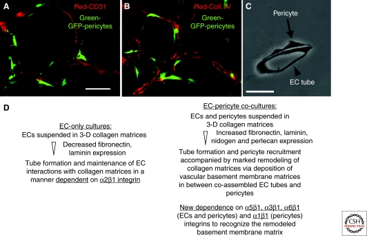
Figure 5.
Pericyte recruitment to EC-lined tubes leads to vascular basement membrane matrix assembly in 3-D matrices. Green-fluorescent protein (GFP)-tagged bovine retinal pericytes were mixed with ECs in 3-D collagen matrices over a period of 5 days, during which EC tubes formed and recruited pericytes. Cultures were fixed at 5 days and immunostained for CD31 (panel A) or for collagen type IV (panel B); bar equals 50 µm. Thin plastic sections were obtained and photographed under transmitted light (panel C); bar equals 25 µm. (Panel D) RT-PCR and protein analyses of EC-only and EC-pericyte cocultures revealed specific changes in ECM and integrin expression, indicating distinctly different regulation in EC-pericyte cocultures in comparison with ECs alone (Stratman et al. 2009a).

