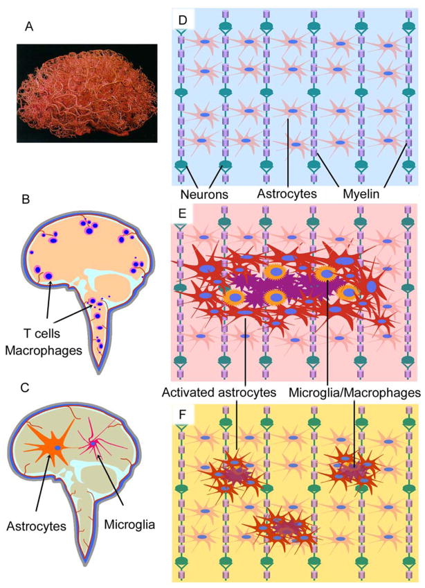
Figure 2. CNS vasculature and inflammation in neurological diseases.
A. Vasculature in the brain. To reveal the vascular web, the brain was injected with a plastic emulsion and the parenchymal tissue was dissolved subsequently. The brain capillaries form a web that encloses the neural networks, supplying CNS nutrients and taking away unneeded wastes (Permission obtained from Wolters Kluwer Health C. Zlokovic, B. V. & Apuzzo, M. L. J. Strategies to circumvent vascular barriers of the central nervous system. Neurosurgery 43(4), 877–878, 1998.). B-F. Schematic drawing shows the CNS in normal or different disease conditions. Two types of inflammation are proposed to contribute to CNS diseases. One is characterized by infiltrating T cells and macrophages (B), which are mainly observed in classical inflammatory disorders such as MS, whereas the other is featured by activation of astrocytes and microglia (C) that is the typical pathological change of the degenerative CNS diseases such as AD, PD and ALS. (D) Neural cells in the CNS form network structures to support each other, in which oligodendrocytes ensheath the axon fibers and astrocytes form nonoverlapping domains. In response to CNS injury, astrocytes get activated with hypertrophy and seal the lesions by forming glial scar (E) or by inducing mild astrogliosis (F). Note that normal astroglial domains are interrupted by astrogliosis, which effects neuron functions.

