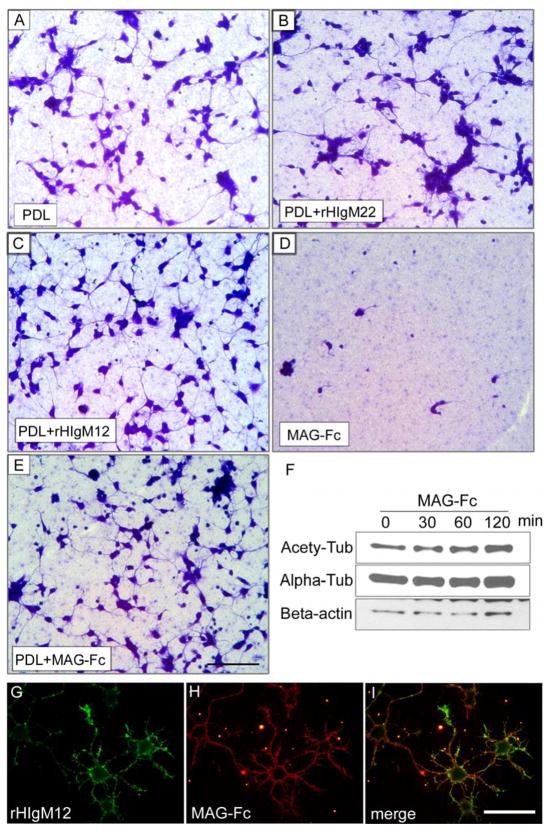
Figure 3. MAG supports neuron stability.
A–E. Primary hippocampal neurons were seeded on nitrocellulose-attached 24-well plates pre-coated with different substrates. The cultures were fixed and stained with Coomassie blue to show the attached neurons 16 hours after plating. Note that neurons grew on myelin-associated glycoprotein fused with human immunoglobulin Fc fragment (MAG-Fc) substrates (D) did not attach well and showed shorter neurites, the floating cells were washed away after fixation. When poly-D-lysine (PDL) plus MAG-Fc were used as substrates (E), MAG-Fc did not inhibit neurite extension as compared with PDL alone (A) or PDL+rHIgM22 (B). HIgM12, which was shown to promote neurite outgrowth[114], was used as positive control (C). F. DIV3 hippocampal neurons were treated with 2.5 μg/ml of MAG-Fc at different times. The level of acetylated tubulin increased after 60 min of treatment, indicating a change in microtubule stability. G–I. DIV1 live hippocampal neurons were incubated with 100 μg/ml of MAG-Fc for 30 min on ice and then stained with rHIgM12. MAG-Fc did not block rHIgM12 binding, and neither did rHIgM12 block MAG-Fc binding (data now shown). Scale bar 50 μm.

