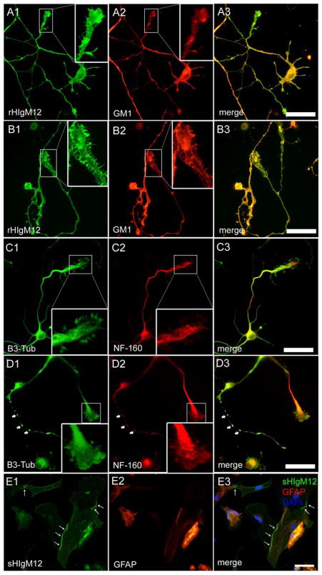
Figure 4. rHIgM12 binds neural cells in different patterns.
A–D. DIV3 hippocampal neurons treated with (B & D) or without (A & C) 100 nM H2O2 were stained with rHIgM12 or anti-cytoskeletal protein antibodies after fixation. Note that rHIgM12 bound both healthy and injured neuronal membranes (not permeabilized) in similar but distinct patterns. However, H2O2 treatment changed cytoskeletal organization substantially. In H2O2-treated neurons, varicosity structures containing both beta-3-tubulin (B3-Tub) and neurofilament 160 (NF-160) were formed along some neurites (arrow head), and cytoskeletons were depolymerized (see high power growth cone regions). E. Compared to the even distribution on neurons, rHIgM12 staining (E1) was enriched along the cell-cell contact in some astrocytes (E2) indicating a distinct binding pattern. Scale bar 50 μm.

