Figure 2.
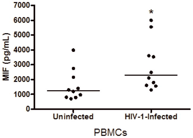
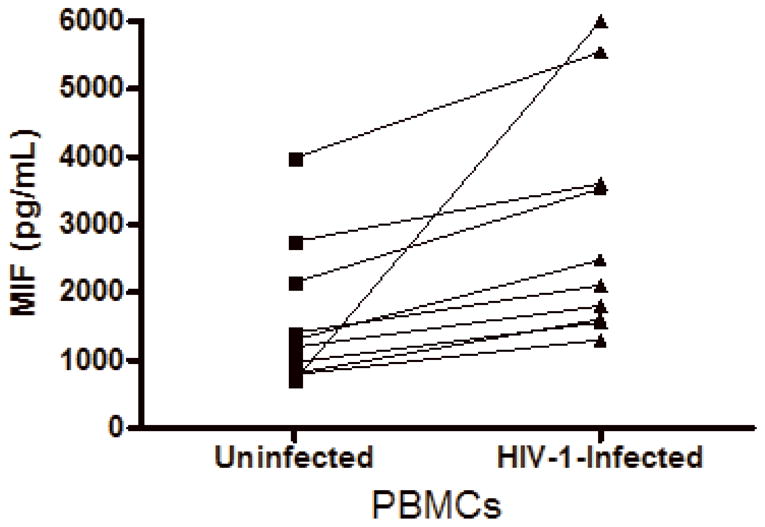
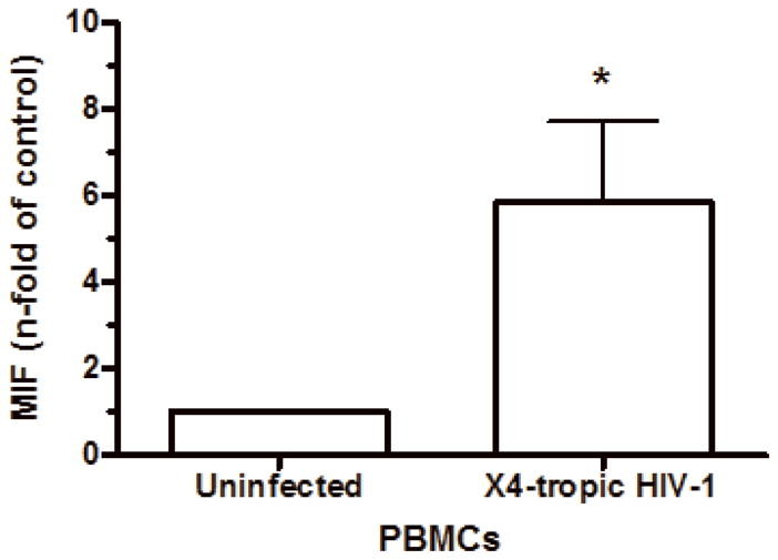
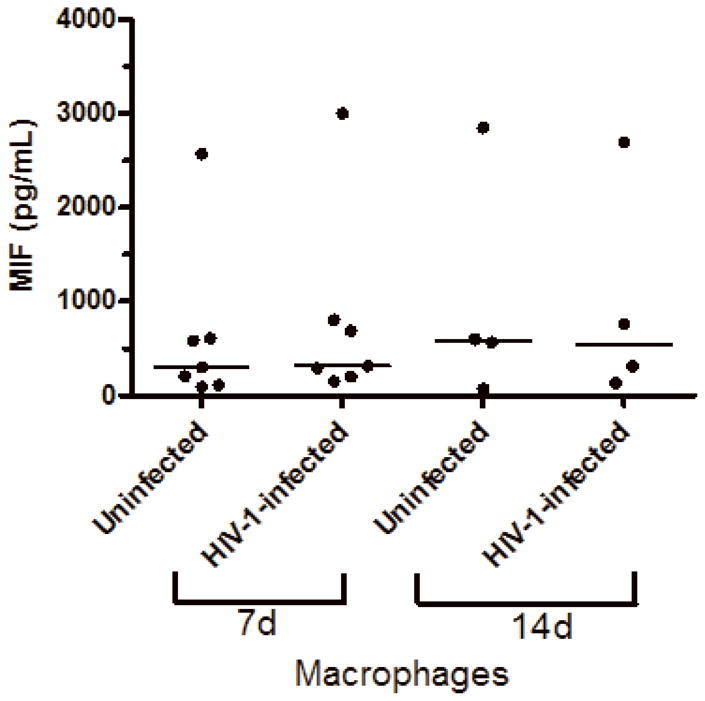
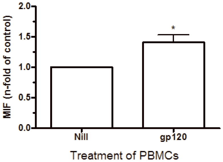
HIV-1 infection and HIV-1 gp120 induce MIF release by PBMCs. (A) PBMCs were infected by an R5-tropic HIV-1 isolate (Ba-L), and after 7 days MIF contents were evaluated in the culture supernatants by ELISA (* p<0.04). Individual values and medians are represented for each group. (B) Lines show individual MIF release in the uninfected and in HIV-1-infected PBMC depicted in figure 2-A. (C) MIF contents in culture supernatants of PBMCs infected by an X4-tropic HIV-1 isolate (Tybe) (* p<0.04). Mean of MIF values by cells cultured only with medium (Nill): 757.8 pg/mL. (D) Macrophages were infected by the HIV-1 isolate Ba-L and MIF release was assessed by ELISA at 7 (7d) and 14 (14d) days after infection. Individual values and medians are represented for each group. (E) Uninfected PBMCs were exposed to recombinant HIV-1 envelope protein gp120 (5 μg/mL) derived from the HIV-1 isolate Ba-L, and MIF secretion was evaluated in culture supernatants by ELISA after 16 hours (* p<0.006). Mean of MIF values by cells cultured only with medium (Nill): 427 pg/mL. In (C) and (E) bars show means ± SEM for 3 and 5 different donors, respectively, and all experiments were done in triplicates per donor.
