Figure 5.
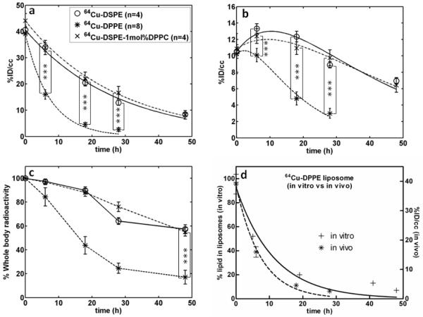
Quantitative analysis (%ID/cc) of PET images with the time activity curve (TAC) of (a) blood pool and (b) liver and (c) whole body radioactivity. Three liposomes (64Cu-DSPE liposomes, 64Cu-DPPE liposomes, 64Cu-DSPE-1mol%DPPC liposomes) were applied. (d) Comparison of stability of 64Cu-DPPE liposomes from blood pool (in vivo) and serum incubation (in vitro) (curve fitted with one-phase exponential decay). Statistical analysis was performed by ANOVA, followed by Tukey’s multiple comparison test (error bars, mean ± STD; ***, P < 0.001)

