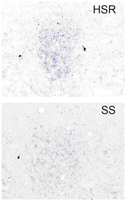
Figure 10.
Photomicrographs of UCN1-positive fibers in the midbrain, rostral to the dorsal raphe nucleus, in a representative highly stress-resilient (HSR) animal and a stress-sensitive (SS) animal. Sections were immunostained for UCN1 and segmented into positive- and negative stained pixels. Positive pixels are highlighted in blue. Cell bodies or debris were erased prior to pixel quantitation. Visually, there were few UCN1 fibers in the SS groups. From (Weissheimer et al., 2010).

