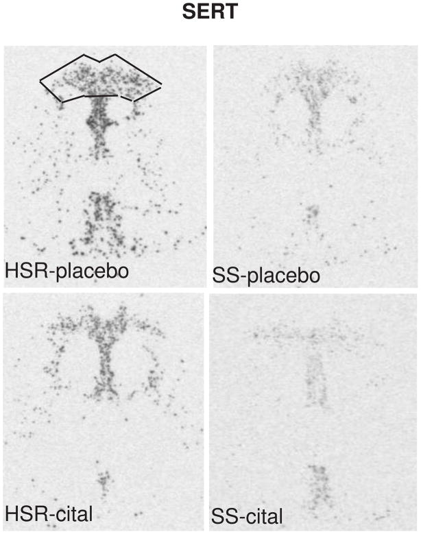
Figure 5.
In situ hybridization autoradiograms of SERT signal in the dorsal raphe nucleus of representative HSR and SS macaques treated with placebo or citalopram for 15 weeks. Scattered neurons of the rostral median raphe are also visible underneath the decussation of the cerebellar peduncles. The box illustrates the area analyzed at this morphological level. The same box was applied to all sections at this level. Each of the 7 levels had a different sized box that was held constant for all of the sections at that morphological level. From (Lima et al., 2009).

