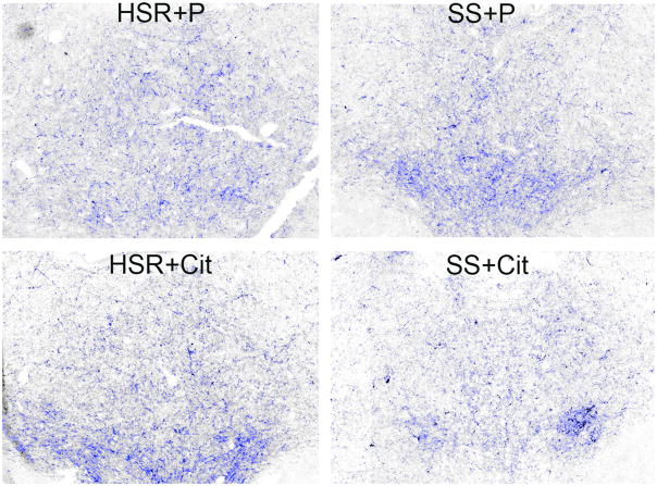
Figure 8.
Photomicrographs of CRF fiber staining in the dorsal raphe nucleus. Stereology montage (×10) of CRF fiber staining in the dorsal raphe nucleus in a representative animal from each treatment group is as follows: HSR+P - Highly stress resilient treated with placebo; HSR+Cit- highly stress resilient treated with s-citalopram; SS+P - stress sensitive treated with placebo; SS+Cit - stress sensitive treated with s-citalopram. Sections were immunostained for CRF and segmented into positive- and negative stained pixels. Positive pixels are highlighted in blue. Cell bodies or debris were erased prior to pixel quantitation. Visually, there appeared to be a lower density of CRF fibers in the SS+Cit treated animal. From (Weissheimer et al., 2010).

