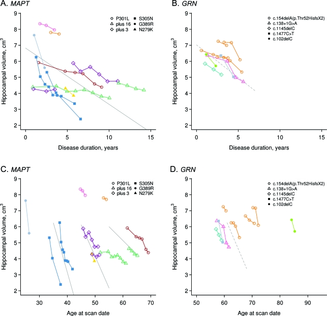
Figure 2. Trajectories of hippocampal volume loss in GRN and MAPT.
(A, B) Hippocampal volume plotted against disease duration. (C, D) Hippocampal volume plotted against age at scan. Data points for individual subjects are shown with the different colors representing different families. The legend highlights the specific mutations of each subject. Volume estimates from 3 T scans are adjusted downward by 0.036 cm3 to remove slight field-strength effects. The solid line in A represents the average volume as a function of disease duration for MAPT subjects assuming age at baseline of 49 years, disease duration at baseline of 1.6 years, and total intracranial volume (TIV) of 1.44 L, the median values in the group. The dashed line in B represents the average volume for GRN subjects assuming age at baseline of 61 years, duration at baseline of 1.9 years, and TIV of 1.40 L, the median values in the group. The solid lines in C contrast average volume for MAPT subjects as a function of age comparing subjects with baseline ages of 35, 45, and 55 years, assuming duration at baseline of 1.6 years and TIV of 1.44 L. The dashed line in D represents average volume for GRN subjects as a function of age assuming duration at baseline of 1.9 years and TIV of 1.40 L.

