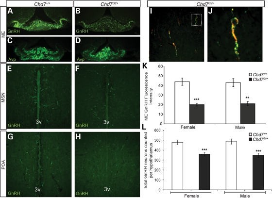
Figure 3.
Chd7Gt/+ mice have decreased GnRH neurons in the hypothalamus. (A and B) Immunofluorescence using anti-GnRH (green) revealed decreased GnRH immunofluorescence in the median eminence in Chd7Gt/+ mice compared with wild-type littermates. (C and D) Immunofluorescence using anti-AVP showed no difference in the median eminence in Chd7Gt/+ mice compared with wild-type littermates. (E–H) Immunofluorescence using anti-GnRH (green) showed decreased numbers of GnRH neurons in the MSN and POA in Chd7Gt/+ mice compared with wild-type littermates. (I and J) Chd7 is expressed in GnRH neurons in vivo. Immunofluorescence of Chd7Gt/+ hypothalamic sections using anti-β-galactosidase (β-gal) and anti-GnRH showed that most GnRH-positive neurons in the hypothalamus are also positive for the β-galactosidase reporter for Chd7 expression. White box in (I) indicates the co-labeled neuron magnified in (J). (K) Quantitation of anti-GnRH immunofluorescence in the median eminence using ImageJ software showed significantly decreased GnRH in Chd7Gt/+ mice compared with the wild-type. (L) Quantitation of GnRH neurons per hypothalamus showed decreased numbers of GnRH neurons in both male and female Chd7Gt/+ mice compared with sex-matched wild-type littermates. Sections are in the coronal plane. **P < 0.01, ***P < 0.001 by unpaired Student's t-test. 3v, third ventricle.

