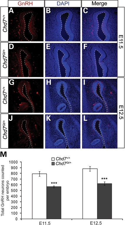
Figure 4.
GnRH neurons are reduced in Chd7Gt/+ embryos. (A–L) Immunofluorescence using antibody against GnRH (red) and counterstained with DAPI (blue) showed a decrease in anti-GnRH staining at E11.5 (A–D) and E12.5. (M) The total number of GnRH neurons per embryo was significantly decreased at each time point in Chd7Gt/+ embryos compared with wild-type littermates. Sections are in the transverse plane. ***P < 0.001 by unpaired Student's t-test.

