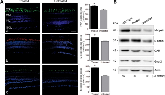
Figure 8.
Gene therapy improves cone survival in Cngb3−/− mice. Injections were performed at P15 and eyes were collected at 60 days post-injection for PNA lectin cytochemistry and analyses of cone-specific protein expression. (A) PNA labeling and cone-opsin staining in the treated Cngb3−/− eyes, compared with the untreated control eyes. Retinal sections were analyzed for: (a) PNA lectin staining, (b) M-opsin and (c) S-opsin immunostaining. Shown are representative images of the analysis from 4 to 6 mice/group and correlative quantitative results. OS, outer segment; ONL, outer nuclear layer; INL, inner nuclear layer, GCL, ganglion cell layer. Scale bar: 50 μm. Quantitative results were obtained from 14 to 18 retinal sections prepared from four mice in each group. Unpaired Student's t-test was used for determination of the significance (P < 0.01). (B) Expression of M-opsin, S-opsin, CAR and GNAT2 in the injected-eyes analyzed by western blot analysis. Retinal membrane preparations were resolved by 10% SDS–PAGE, followed by immunoblotting using antibodies against the respective proteins. Shown are representative images of the analysis from 6 to 8 mice/group.

