Figure 5.
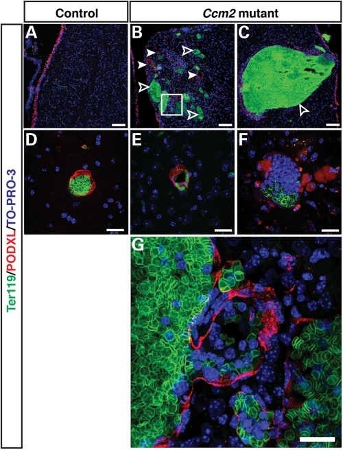
Vessel dilation and lumen breakdown in the cerebrum of the inducibly deleted, adult Ccm2 mutant mice. Triple immunofluorescence confocal microscopy of coronal sections was performed with antibodies to a vascular lumen marker Podocarxyn (PODXL) (red), together with the erythrocyte marker Ter119 (green) and the pan-nuclear marker TO-PRO-3 (blue). The pIpC-treated Ccm2flox/flox;MX1-Cre mice have dilated vessels (B, arrowheads), and damaged vessels which failed to form a proper vascular lumen (B, open arrowhead; C and F) in hemorrhaged areas (CCM-like lesions),while the pIpC-treated control littermates have only normal-sized vessels with well-demarcated lumen (A and D). Normal capillaries are also present in Ccm2flox/flox;MX1-Cre mice in areas that do not show any hemorrhage (E). The boxed region in (B) is shown at higher magnification in (G). The vascular lumen appears to have ruptured resulting in hemorrhage, as evidenced by erythrocytes outside the vessel enclosure. Scale bars: A, B, C are 100 µm; D, E, F, G are 20 µm.

