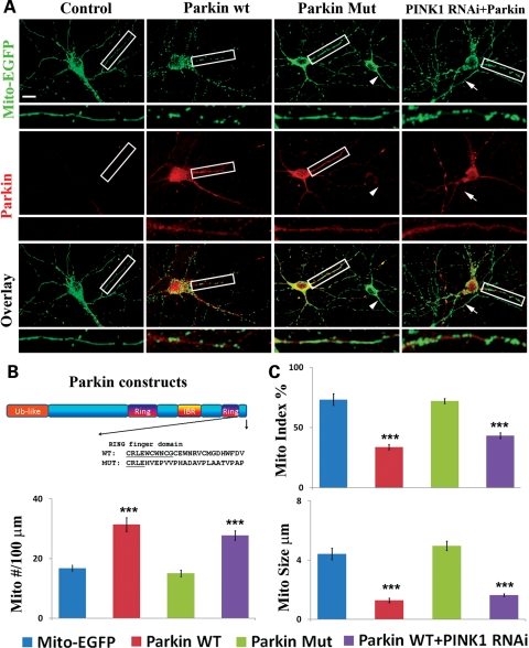
Figure 2.
Parkin overexpression leads to mitochondrial fragmentation and it suppresses PINK1 RNAi effect. (A) Cultured hippocampal neurons were transfected with Mito-EGFP and myc-tagged Parkin-wt, Parkin-mut or Parkin-wt plus PINK1 RNAi. Immunofluorescence was performed with chicken anti-EGFP and mouse anti-myc antibodies. Neurons marked with arrowheads in the Parkin-mut panels are control neurons transfected with Mito-EGFP only. In the PINK1 RNAi + Parkin panels, neurons were transfected with the lentiviral vector expressing PINK1 shRNA, mito-EGFP and myc-tagged Parkin-wt. Arrows show mitochondrial morphology in a neuron transfected with PINK1 shRNA only. (B) A diagram showing the human Parkin constructs used. (C) Quantification of Mito index, Mito number and Mito size in the dendrites of neurons transfected with the constructs is shown in (A)(***P< 0.001 in Student's t-test).

