
Fig. 3.
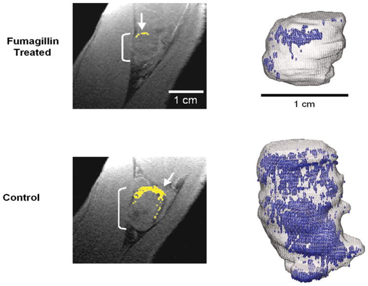
Diminished αvβ3 integrin contrast enhancement in T1-weighted, fat-suppressed, 3D gradient echo MR single slice images (250 ×250 μm, 500 μm slices, TR/TE = 40/5.6 ms, 65° flip angle, 1.5 T) in rabbits administered αvβ3-targeted fumagillin nanoparticles (top) versus those given αvβ3-targeted nanoparticles without drug (bottom). Left: Enhancing pixels, color coded in yellow (arrows), demonstrate sparse areas of angiogenesis in fumagillin-treated animal (top). Right: 3D neovascular maps of example Vx-2 tumors on day 16 following αvβ3-targeted fumagillin nanoparticles (top) versus αvβ3-nanoparticles without drug (bottom). Note the asymmetric distribution of angiogenic signal (color coded in blue) over the tumor surface in both the control and treated animals. Neovessel dense islands and the interspersed fine network of angiogenic proliferation over the tumor surface are diminished in rabbits receiving the targeted fumagillin treatment. Reproduced with permission [63]