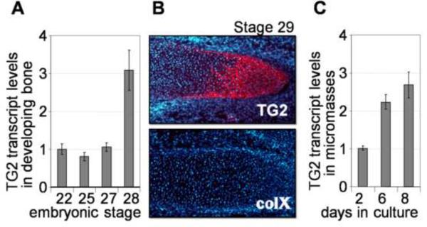
Figure 1.
TG2 expression in the developing chicken limbs. A –levels of TG2 mRNA in mesenchymal cells isolated from the limb buds or cartilaginous anlagen at stages 21–28 of embryonic development, analyzed by real-time RT-PCR. B – immunological detection of the TG2 protein (red, upper panel) in the prehypertrophic cartilaginous anlage of a day 6.5 (stage 29) embryo. The same anlage is negative for collagen type X protein (as indicated by the lack of red signal on the lower panel). Cell nuclei are counterstained with DAPI (blue). C –real-time RT-PCR analysis shows up-regulation of the TG2 mRNA during chondrogenic differentiation of micromass cultures (normalized to beta-actin expression).

