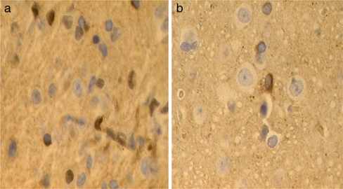
Fig. 3.
The c-fos staining in the frontal lobe of IBS model and control. The c-fos staining can be seen both in cytoplasm and nucleus. The yellow-brown c-fos positive neurons distribute irregularly in frontal lobe. The verge of cell is distinct. The morphology of cyton can be spindle-shaped, oval, or irregular. a c-fos in frontal lobe of IBS model (×400). b c-fos in frontal lobe of control (×400)

