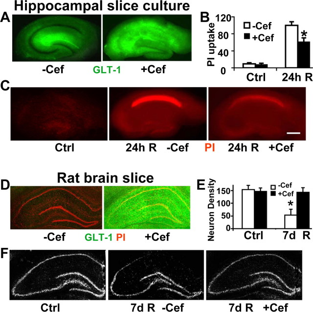
Figure 5.
Ceftriaxone induces GLT-1 expression and reduces CA1 delayed neuronal injury in hippocampal slice culture and in vivo. A, Ceftriaxone pretreatment (+Cef; 100 μm for 5 d) strongly induces expression of GLT-1 immunoreactivity (green) in hippocampal slice cultures, especially in the CA1 subregion, compared with the vehicle-treated control (−Cef). B, Bar graph shows the significant reduction of CA1 neuronal damage in ceftriaxone-treated (+Cef) slices compared with vehicle-treated (−Cef) ones by relative PI fluorescence in hippocampal slice culture 24 h after 15 min OGD. n = 9 slices per condition; *p < 0.05 indicates significant difference from control. C, Fluorescence images of representative PI-stained (red) hippocampal slices 24 h after 15 min OGD. Decreased PI fluorescence indicates decreased CA1 neuronal death in ceftriaxone-treated (+Cef) slices compared with vehicle-treated (−Cef) ones. Scale bar, 400 μm. D, Pretreatment of rats with ceftriaxone (200 mg/kg, i.p., daily for 5 d) strongly induces expression of GLT-1 throughout the hippocampus. E, Bar graph shows the reduction of CA1 neuronal damage in ceftriaxone-treated (+Cef) animals compared with vehicle treated (−Cef) rats by cresyl violet staining intensity. n = 4 in each group; *p < 0.05 significantly different from control. F, Cresyl violet staining of representative brain slices from control and ischemic rats pretreated with ceftriaxone (+Cef) or vehicle (−Cef). More pyramidal neurons were present in ceftriaxone-treated brains after cresyl violet staining compared with those in the vehicle-treated group after 7 d reperfusion. Ctrl, Control group; R, reperfusion.

