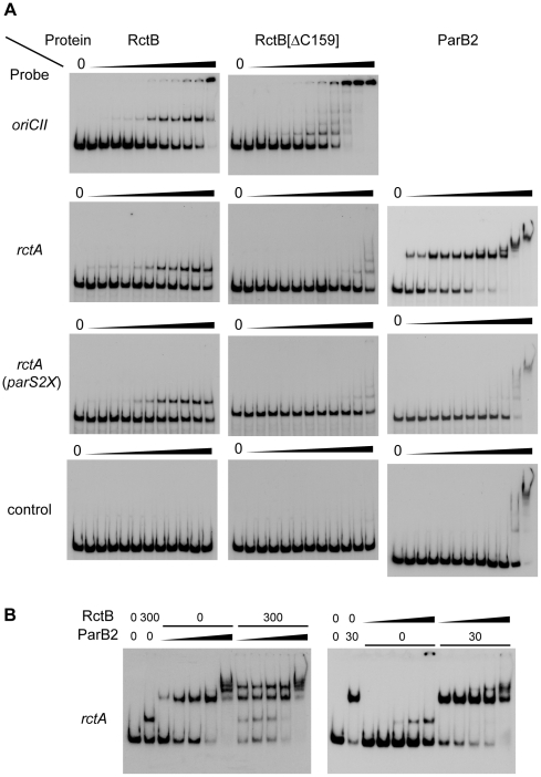
Figure 3. Binding of RctB and/or ParB2 to DNA fragments containing rctA and parS2.
A) Binding of wild type or mutant RctB or ParB2 proteins to indicated DNA fragments. The amount of protein used in each lane was 0, 0.01, 0.03, 0.1, 0.3, 1, 3, 10, 31, 100, 316, 1000 ng, from left to right. B) Binding of RctB and ParB2 proteins to rctA. RctB and ParB2 were premixed and then added to the reaction tube. Amounts of proteins (ng) are indicated and the dilution series from left to right was 0.03, 0.3, 3, 30 and 300 ng. Note that similar banding patterns were observed in experiments where either RctB or ParB2 was added prior to the other protein (see Figure S1).

