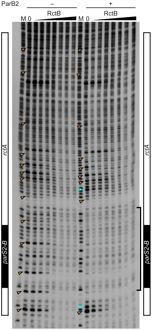
Figure 4. Protection of rctA from DNase I digestion in the presence of RctB or RctB and ParB2.
The DNase I protection assay was performed with 0, 10, 20, 40, 80, 160, 320 and 640 ng RctB bound to a 5′-32P-labeled DNA probe containing rctA (including parS2-B, indicated at the side of the gel), in the absence or presence of ParB2 (100 ng). M denotes the G+A chemical sequencing ladder. The bracket indicates the ParB2 footprint. The arrowheads indicate nucleotides protected by RctB in multiple independent experiments. The arrows indicate hypersensitive sites.

