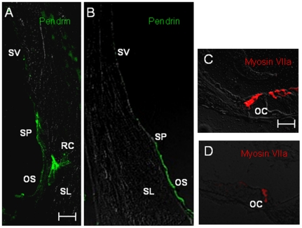
Figure 2. Immunolocalization of pendrin in Slc26a4+/+ mice (A) and Slc26a4tm1Dontuh/tm1Dontuh mice (B).
The expression of pendrin (stained in green) in spiral prominence and outer sulcus epithelial cells was not affected by the c.919-2A>G mutation. Degeneration of cochlear hair cells in Slc26a4tm1Dontuh/tm1Dontuh mice was revealed by fluorescence confocal microscopy (D). (OC, organ of Corti; OS, outer sulcus epithelial cells; RC, root cells; SP, spiral prominence; SL, spiral ligament; SV, stria vascularis). Bar = 50 µm.

