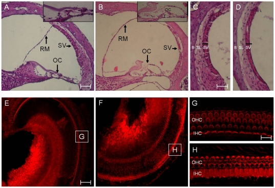
Figure 5. Comparison of cochlear morphology between Slc26a4+/+ mice (A, C, E & G) and Slc26a4tm1Dontuh/tm1Dontuh mice (B, D, F & H).
Microscopic examination of the cochlear duct revealed severe endolymphatic hydrops with dilatation of scala media, degeneration of hair cells (B) and a significant atrophy of the stria vascularis (D) in Slc26a4tm1Dontuh/tm1Dontuh mice. Degeneration of cochlear hair cells in Slc26a4tm1Dontuh/tm1Dontuh mice was also revealed by fluorescence confocal microscopy (H). G, H: magnification of boxes G and H from figures E and F, respectively. B, bone; IHC, inner hair cells; OC, organ of Corti; OHC, outer hair cells; RM, Reissner's membrane; SL, spiral ligament; SV, stria vascularis; T, tunnel of Corti. Bar = 150 µm (A & B), 50 µm (C, D, E & F), and 10 µm (G & H).

