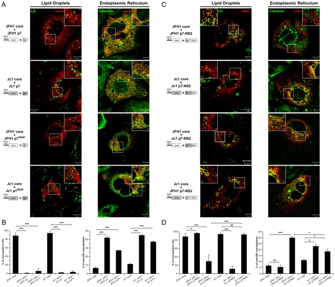
Figure 6. Strain-specific influence of p7 and NS2 on the intracellular localization of core.
Huh7.5 cells stably expressing p7 or mutated p7, JFH1-p7mut and Jc1-p7mut (RR33/35AA JFH1-p7 or KR33/35AA Jc1-p7 respectively) proteins (A, B) and p7-NS2 (C, D) from JFH1 and J6-CF (Jc1) isolates were transfected with plasmids expressing core from the same HCV strain or from the other isolate. 72 h post-transfection, cells were stained for LDs, Calnexin, and HCV core proteins. Intracellular localization of core proteins (red channel) in LD or ER (green channels) was analyzed by confocal microscopy. Typical patterns of intracellular localization of either protein are shown. The scale bars are provided in each panel as well as in zooms from squared areas. The constructs expressed in transfected cells are depicted above each panel (A, C). The frequency of core-positive LDs (mean % ± SD) was determined in HCVcc-containing cells stained for core and LDs (left panel). The percentages of core-ER colocalization (mean % ± SD) were determined by expressing the coefficients of determination based on Pearson's correlation coefficients of colocalization of core and Calnexin (right panel). For each condition, 30–50 cells were quantified. (*), P<0.05; (**), P<0.01; (***), P<0.001; (ns), no significant difference (B, D).

