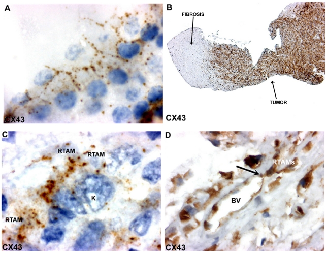
Figure 5. CX43 expression in normal thyroid tissue and in ATC.
A) Normal thyroid tissue: characteristic CX43 positive punctuate gap junctions at the intercellular basolateral membranes of thyrocytes and absence of staining at the apical border. Note a faint diffuse or vesicular staining in the cytoplasm which could correspond to the synthesis and/or transport of the protein to the membrane. Original magnification: ×400. B) Strong CX43 immunostaining in ATC (on the right part of the photo). Non-tumor fibrotic tissue is seen on the left part. Original magnification: ×50. C). Characteristic CX43-positive punctuate junctions localized where the RTAMs and cancer cells (K) come into contact. Original magnification: ×1000. D) CX43 immunostaining of RTAMs and endothelial cells (BV: Blood Vessel). Note a CX43-positive “button” at the junction between the endothelial blood vessel and RTAM (black arrow). Original magnification: ×400.

