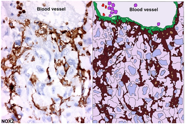
Figure 10. Extensions of interconnected RTAMs from blood vessel to avascular tumor areas.
On the left, in case n°3 in which the number of RTAMs was relatively low and this allowed us to distinguish RTAMs along the blood vessel (B.V.) and the contiguous chain extension of RTAMs from the blood vessel to within the tumor cells. Original magnification: ×200. On the right, a drawing underlining the structure and the relationships between the different components was obtained by copying the photo in transparent digital layer with Adobe Photoshop and adding false colors; RTAMs in brown, cancer cells clear pink and blue; endothelial cells in green, neutrophils and/or monocytes in vessel lumen in violet and lymphocytes in red.

