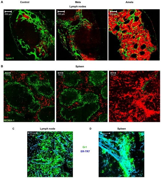
Figure 7. Gr1+CD11b+ cells recruited in SLO interact with the stroma in mice developing Amela-melanomas.
LN (A) or spleen (B) sections from control, Mela- and Amela-bearing mice were stained with anti-Gr1-PE (red) and anti-Lyve-1-biotyl/streptavidin-FITC (green) to detect LN medullas or for MOMA-1 (green) to delineate splenic white pulp areas, respectively. LN (C) or spleen (D) sections from Amela-bearing mice were stained with anti-Gr1-APC (green) and anti-ER-TR7/chicken anti-rat-Alexa647 (blue). Magnification showing a cluster of Gr1+ cells on the stromal network is shown.

