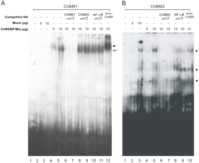
Figure 5. Validation of ChREBP/Mlx binding to two enriched motifs, ChBM1 and ChBM2.
Electrophoretic mobility shift assays were performed with an oligonucleotide containing the ChBM1 (A) or ChBM2 (B) probe. All lanes contain the labeled probe, and lanes 2–12 contain 5 or 10 µg of HEK293 nuclear extract. Lanes 2 and 3 are HEK293 mock-transfected nuclear extract. The other lanes contain extract from HEK293 cells transfected with the ChREBP and Mlx expression plasmids. For competition assays, a 10- or 50-molar excess of various unlabeled competitor DNAs was added to the reaction mixture. Anti-ChREBP (Anti-ChBP, 0.6 µg) was added as indicated. The white arrow indicates the position of the ChREBP/Mlx complex. The black arrow indicates the position of the antibody-supershifted complexes. The asterisks indicate the position of background bands present in the control HEK293 cell nuclear extract.

