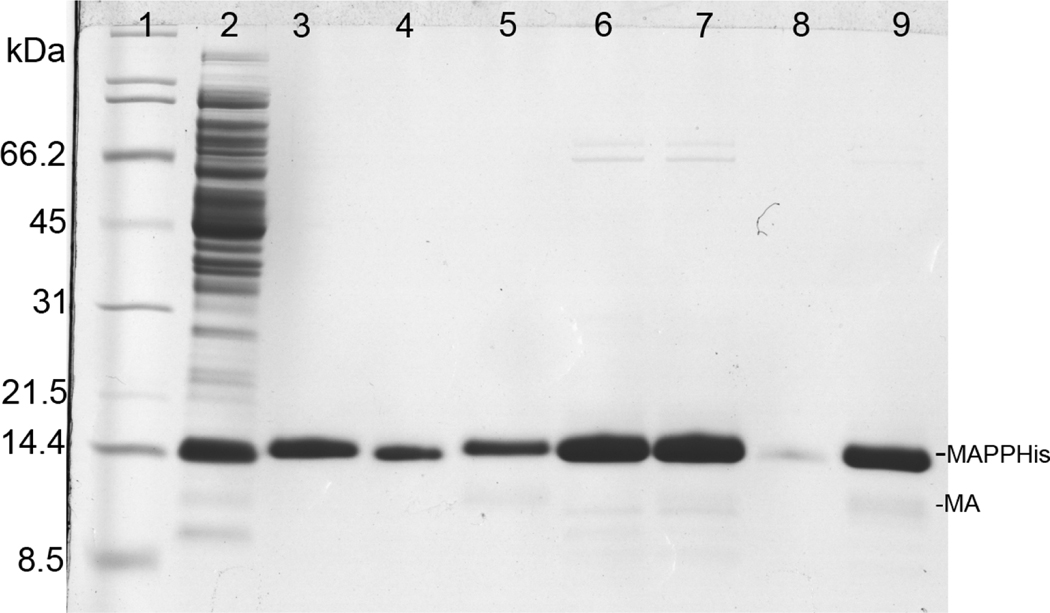
Figure 4.
Coomassie stained SDS-PAGE gel illustrating Ni-NTA purification of myrMAPPHis produced using the two-plasmid system. Lanes 3–5: purification of the first aliquot directly eluted by imidazole buffer and lanes 6–9: purification of the second aliquot, which was cleaved on Ni-NTA and then eluted by imidazole. Lanes: (1) broad range SDS-PAGE standard (Bio-Rad), (2) flow-through after binding (14 kDa band is lysozyme), (3) proteins eluted by imidazole buffer, (4) sample of Ni-NTA agarose after elution, (5) eluate (the same as in lane 3) cleaved by Pr13 for 1 hour, (6) protein cleaved from Ni-NTA by Pr13, (7) protein eluted from column using imidazole buffer after cleaving, (8) sample of Ni-NTA agarose after elution, (9) eluate (the same as in lane 7) cleaved by Pr13 for 1 hour

