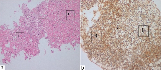
Figure 1.

(a) Low-power view of the liver biopsy core showing preserved hepatocytes around the portal areas (Zone 1) and necrosis in Zones 2 and 3. (b) The reticulin stain showing the preserved framework in Zone 1 and destroyed framework in Zones 2 and 3
