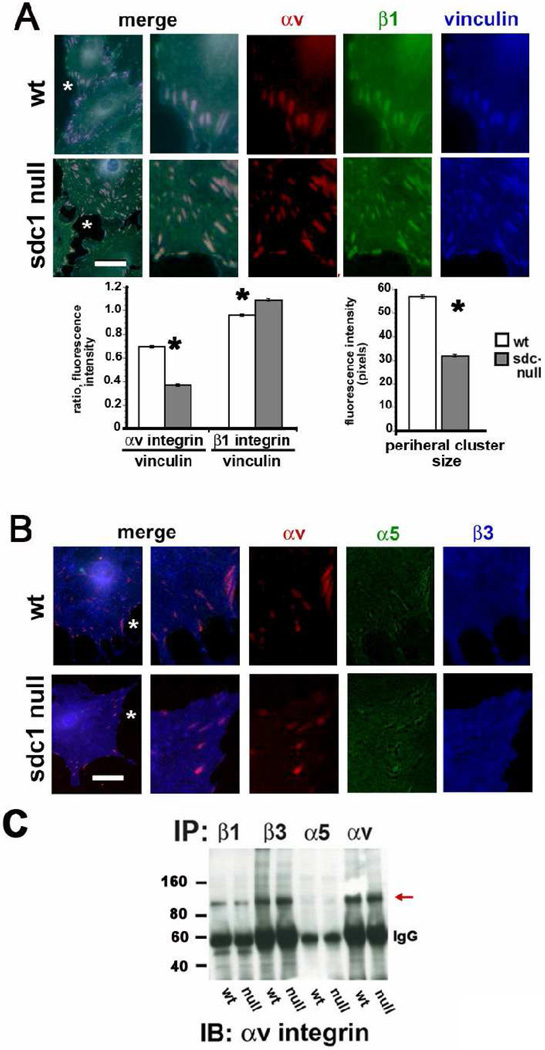
Figure 3. Sdc1 null CSCs assemble focal adhesions containing β1 and αv integrins with less β3 and α5 compared to wt cells.
A and B show wt and sdc1 null cells grown for two days in culture, fixed, permeabilized, and immunostained for vinculin, αv, and β1 integrin, or for vinculin, α5, and β3 integrin respectively. A region of interest identified with an asterisk in each image was digitally enlarged three-fold and presented with the three molecules stained shown individually and merged. Note that β1 and αv integrins co-localize within focal adhesions in both wt and sdc1 null cells and that these same vinculin-containing focal adhesions show little evidence of β3 and α5 integrin. The line tool was used in Image Pro Plus and the ratios of αv and β1 integrins to vinculin determined; also, the mean intensity of the vinculin-containing clusters in pixels in wt and sdc1 null cells. A minimum of ten cells per variable was assessed and no fewer than 100 ratios calculated per cell. Data are shown for the peripheral adhesions in wt and sdc1 null cells. C. Immunoprecipitation of wt and CSC extracts was performed using antibodies against β1, β3, α5 or αv integrins followed by immunoblotting with αv integrin to reveal the presence of αvβ1 integrin heterodimers within CSC extracts. αv is immunoprecipitated using antibodies against β1, β3, and αv integrins but not α5 (red arrow). The asterisks in the bar graphs in A indicate data that are significant by ANOVA with p values < 0.05 and the bar in A and B = 10 µm.

