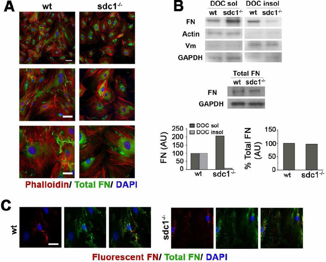
Figure 6. CSCs lacking sdc1 synthesize the same amount of FN but assemble fewer DOC-insoluble FN fibrils.
A. Wild type and sdc1-null CSC were cultured for five days and immunostained with an antibody specific for endogenous FN. Sdc1-null CSCs exhibit fewer endogenous FN fibrils (green) than their wild type counterparts. Staining for F-actin (phalloidin, red) and nuclei (DAPI) are also shown. B. Sdc1 null CSCs exhibit increased DOC soluble and decreased DOC insoluble FN relative to wild type CSCs. Quantification of CSC DOC soluble and insoluble extracts was normalized to levels of β-actin and vimentin, respectively, and expressed relative to wild type CSCs. Vimentin (DOC insoluble) and β-actin (DOC soluble) confirmed separation of the two fractions. In contrast, wild type and sdc1-null CSCs exhibit no difference in the amount of total endogenous FN, with quantification normalized to GAPDH. Shown is one representative experiment of three. C. Sdc1 null CSCs also incorporate less exogenous FN into a fibrillar matrix (red), shown relative to the endogenous matrix (green). The scale bar in A for the top images is 100 µm and is 40 µm for the lower images; in C, the bar is 20 µm. Wt and sdc1 null cells are shown at the same magnification.

