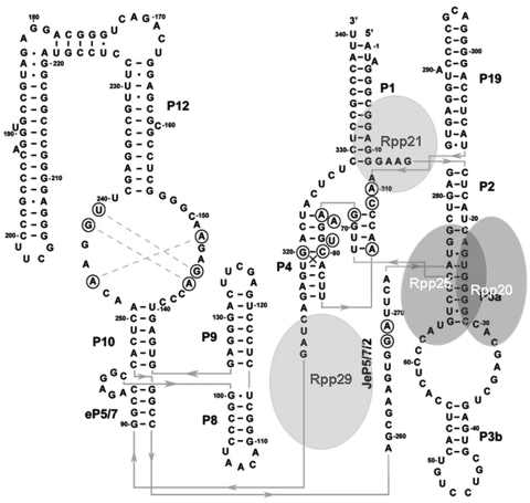
Figure 1.
H1 RNA structure and its interaction with protein subunits. A proposed secondary structure of H1 RNA was deduced from the model of eukaryal RNase P RNA (16) and the crystal structures of bacterial RNase P RNAs (17,18). Four major coaxial helices are seen: P2–P3–P19, P1–P4-JeP5/7/2, P8–P9, eP5/7-P10–P12. The orientation of the extended part of P12 (positions 178–221) relative to the co-axial helix is unknown. Bases in circles are conserved. The approximate binding sites of the protein subunits Rpp20, Rpp25, Rpp21 and Rpp29 in H1 RNA are based on the nuclease footprinting analyses described in this study.

