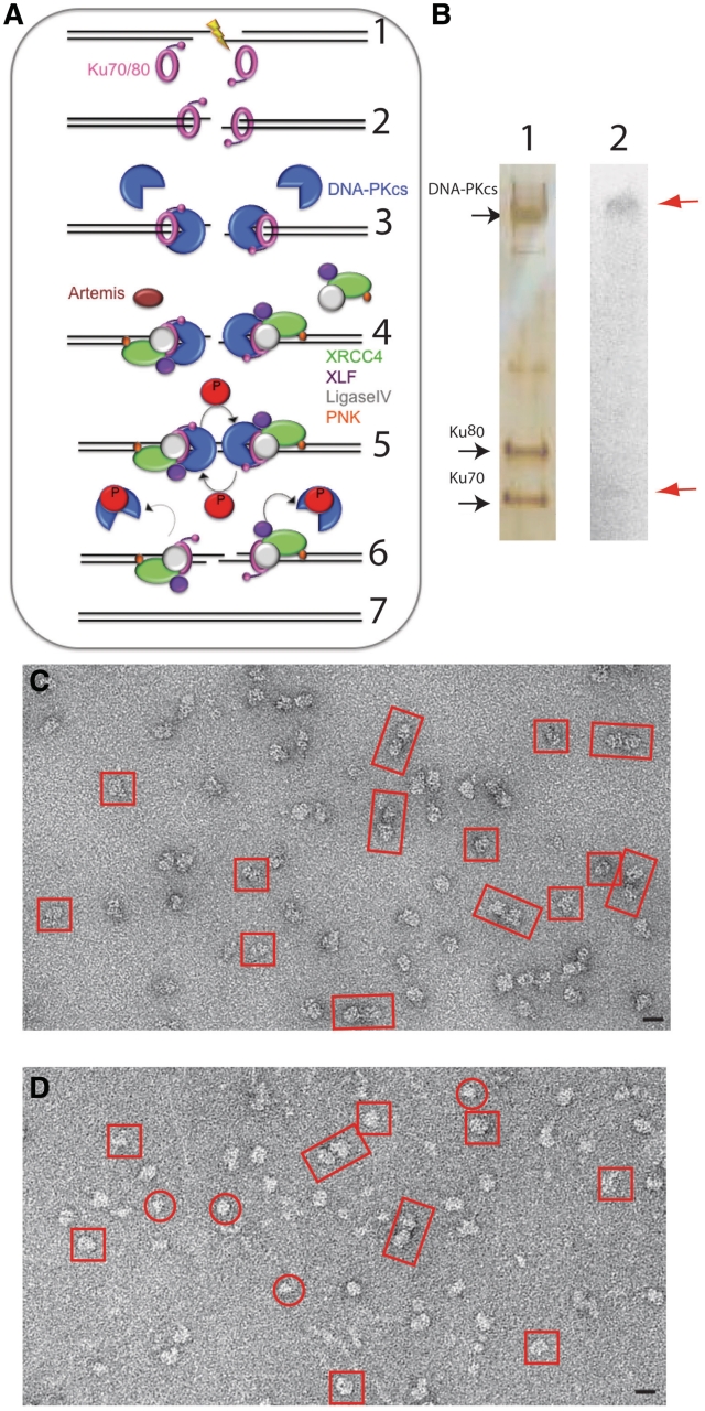
Figure 1.
Effect of autophosphorylation on DNA-PK. (A) A schematic diagram of NHEJ. Ionizing Radiation induces a DSB Step (1). The DSB is detected and bound by the Ku70/80 heterodimer (pink, Step 2). Once bound to the DSB, the Ku80 C-terminal domain recruits DNA-PKcs (blue). Ku translocates inward and positions DNA-PKcs at the extremity of the DSB (Step 3). The DNA ends are processed by one or more possible enzymes that include Artemis (brown), Polynucleotide kinase (PNK, orange), XRCC4 (green), XLF (purple) and LigaseIV (grey) (Step 4). DNA-PKcs undergoes autophosphorylation (indicated by arrows), resulting in the release of autophosphorylated DNA-PKcs from DNA (Step 5). In the final step (Step 6), the XRCC4/DNA ligase IV complex ligates the DNA ends in a reaction that is stimulated by XLF. (B) (1) Silver stained SDS-PAGE gel of autophosphorylated DNA-PK complex, (2) autoradiograph showing autophosphorylation of DNA-PK. The dephosphorylated sample was incubated for 1 h at 37°C in the presence of 10 µM ATP and 0.1 µCi of [γ-32P]ATP in a final volume of 20 µl. Radioactive products were separated on SDS–PAGE on a NuPAGE 4–12% gel and visualized by autoradiography. (C) Electron micrograph of negatively stained dephosphorylated DNA-PK. (D) Electron micrograph of negatively-stained autophosphorylated sample. Dimers are boxed in rectangles, large monomers in squares and small monomers in circles. Scale bar = 200 Å.

