Figure 1.
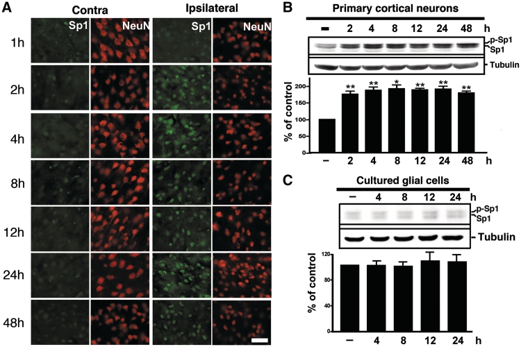
Hypoxia/ischaemia-induced Sp1. (A) Double immunostaining of Sp1 and NeuN at various time points of recovery from MCAO was performed. Brain sections were obtained at various time points after MCAO and were subsequently probed with anti-Sp1 and anti-NeuN antibodies. HIF-1α was detected using Alexa 488 (green) while NeuN was detected using Alexa 568 (red). Panels show images taken from the cortex of non-ischemic and ischemic hemispheres. Scale bar, 50 µm. (B and C) The time course of Sp1 expression induced by OGD treatment in primary neurons or glial cells was assessed. Total lysates of primary neurons (B) or glial cells (C) were obtained at various time points following OGD challenge. Sp1 expression was detected by performing immunoblotting with anti-Sp1 antibodies using tubulin as an internal control. All experiments were independently carried out in triplicate and were expressed as a percentage of the levels in the naïve controls. Statistical analysis was carried out using the one-way ANOVA with appropriate post hoc tests. **P < 0.01 versus the naïve control group.

