Fig. 3.

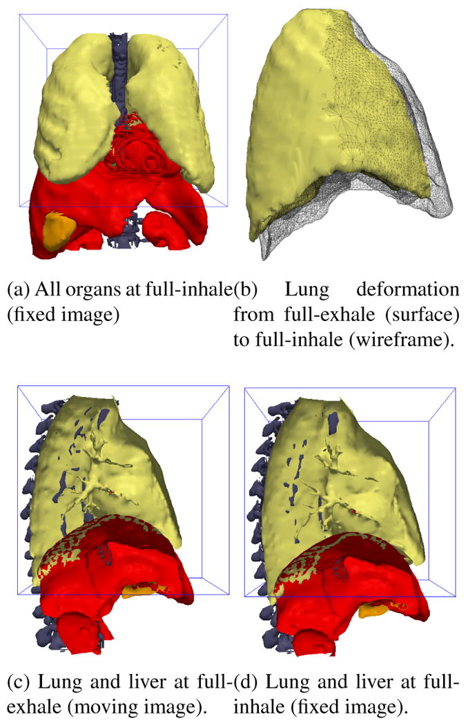
Surfaces extracted from the fixed and moving XCAT phantom images. The 3D box outline indicates the domain of the fixed and moving images to be registered.

Surfaces extracted from the fixed and moving XCAT phantom images. The 3D box outline indicates the domain of the fixed and moving images to be registered.