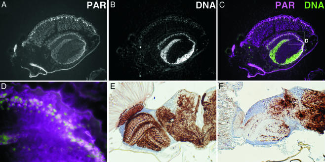
Fig. 3.
Accumulation of poly(ADP-ribose) and disruption of axon structures in the parg mutant. (A) Poly(ADP-ribose) in a parg27.1/Y adult as detected with anti-poly(ADP-ribose) antibody. (B) Nuclei are stained with Hoechst dye 33342. A part of the sagittal section of the head is shown. (C) Merging of poly(ADP-ribose) (magenta) and DNA (green). The box in C is shown in D at higher magnification. Axon staining of the horizontal section of adult head at 10 days after eclosion is shown in E for the wild type and in F for parg27.1/Y mutant.

