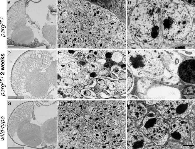
Fig. 4.
Microscopic and electron-microscopic analyses of adult brain. Horizontal section through adult heads. (A–C) parg27.1/Y at a few days after eclosion. (D–F) parg27.1/Y at 2 weeks after eclosion. (G–I) Wild type. A, D, and G show light microphotographs of tissue stained with toluidine blue; B, E, and H show electron microphotographs; C, F, and I show higher magnification of parts of B, E, and H, respectively. Open arrowheads indicate granular structures typical in the parg27.1/Y genotype. (Scale bars, 2 μm in B, E, and H and 1 μm in C, F, and I.)

