
Figure 6.
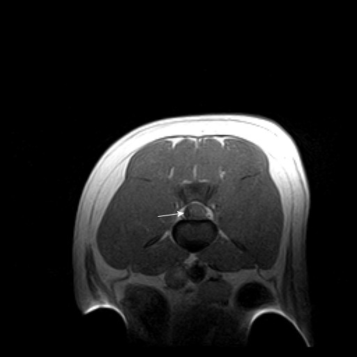
Magnetic resonance imaging of the lumbar spinal cord, case 4. Transverse T1 weighted images of the spinal cord at the level of vertebrae L5 revealing a hypointense area (arrow) lateralised to the right and compressing the spinal cord towards the left.