Figure 7.

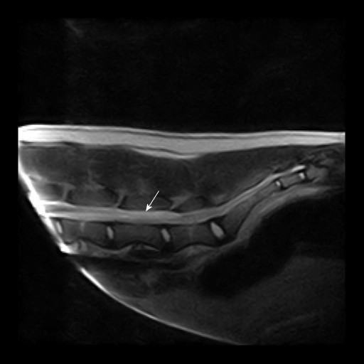
Magnetic resonance imaging of the lumbosacral area, case 4. Sagital T2 weighted images of the lumbosacral spinal cord demonstrating a hyperintense area (arrow) compatible with oedema caudal to the suspected haemorrhage.

Magnetic resonance imaging of the lumbosacral area, case 4. Sagital T2 weighted images of the lumbosacral spinal cord demonstrating a hyperintense area (arrow) compatible with oedema caudal to the suspected haemorrhage.