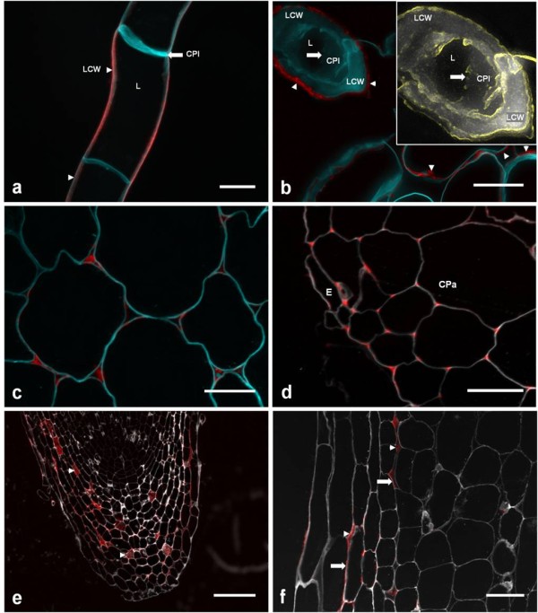
Figure 7.

JIM11 detection of extensin epitopes in selected tissues of grapevine, potato and Arabidopsis. A) Confocal image of frozen callus. B) 0.5 μm sections of resin-fixed callus. Inset: Magnified image of calcofluor signal from transverse section of callus cell (from top left corner), in which edge detection (yellow) was used to highlight the spatial limit of the broken cell plate. N.B. JIM11 signals are in lateral cell walls (arrowheads), and not the cell plate (arrows). C) Cortical parenchyma of basal grapevine epicotyl. D) Epidermal and cortical parenchyma of root. E) Root cap. Note the presence of JIM11 epitopes in large intercellular spaces (arrow heads). F) Higher amplification of lateral outer layer of root cap. N.B. JIM11 epitopes are present in both intercellular spaces (arrow heads) and cell wall (arrows). Scalebars: A-F, 25 μm; G, 250 μm; H, 100 μm. In all cases, calcofluor (for cellulose marking) signals were false-coloured to cyan (panels (A-E) or white (F-H). Key: LCW, lateral cell wall; CPl, cell plate; L, lumen; E, epidermis; CPa, cortical parenchyma;
