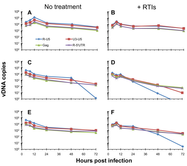
Figure 6.
NC mutant reverse transcription kinetics in cells are altered when premature reverse transcription is blocked. CD4+ HeLa cells were infected with virus prepared in the absence or presence of RTIs that were subsequently removed using PEG-precipitation (Figure 1, left). These charts display the profile of reverse transcripts over a 72 h time course of infection. Panels A, C, and E show infections from viruses (WT, NCH23C, and NCH44C, respectively) not treated with RTIs, and panels B, D, and F show infections from viruses (WT, NCH23C, and NCH44C, respectively) where premature reverse transcription was blocked via RTI treatment. Prior to the infection, all of the virus samples were normalized for RT activity so that equal amounts were used to infect each set of cells. These results are from a representative experiment. The vDNA species measured were normalized for cell equivalents using CCR5 and are indicated at the bottom of panel A. Schematics of the pertinent vDNA target sites are shown at the bottom of Figure 3.

