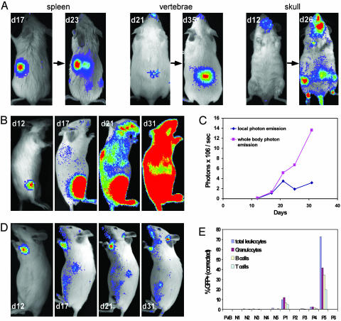
Fig. 4.
Single HSC engraftment. Single luc+ or luc+ GFP+ KTLS HSC were injected i.v. into lethally irradiated FVB recipients along with 3 × 10⁵ Sca-1-depleted BM cells as a radioprotective population. All images are displayed at the same scale. (A) Representative images showing single HSC-engrafted mice in which single foci from spleen, vertebrae, and skull were observed. These foci expanded with time, and new detectable sites became apparent at later time points. (B) Right lateral view images were taken on days 12, 17, 21, and 31 from one animal in which one initial bioluminescent focus was visualized on day 12 after transplantation. By day 31, a significant degree of hematopoietic reconstitution was apparent. (C) Kinetics of hematopoietic reconstitution of the recipient shown in B showed steady increases in whole body bioluminescent emission with relatively stable signal detected from the initial focus (local photon emission). (D) One focus from another single HSC recipient yielded only low-level hematopoietic engraftment. Left lateral view images were taken on days 12, 17, 21, and 31. (E) Chimerism evaluation of peripheral blood of the single luc+ GFP+ HSC recipients in a representative experiment, including wild-type FVB and recipients with (P1–6) and without (N1–5) initial foci. At week 5, 72.7% of hematopoietic cells from P5 (recipient shown in B) were derived from donor HSC, whereas only 0.3% of those from P6 (recipient shown in D) were derived from the labeled donor cell.

