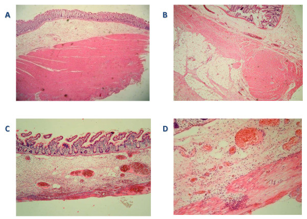
Figure 3.
Several microscopic histological slides obtained from the patient. (A) A segment of normal colon from this patient. (B) A segment of abnormal colon and (C) a segment of abnormal small bowel showing patchy thinning and fibrosis of the muscularis externa. (D) Abnormal muscle with patchy fibrosis (sigmoid colon). It is also inflamed because of the perforation in the region.

