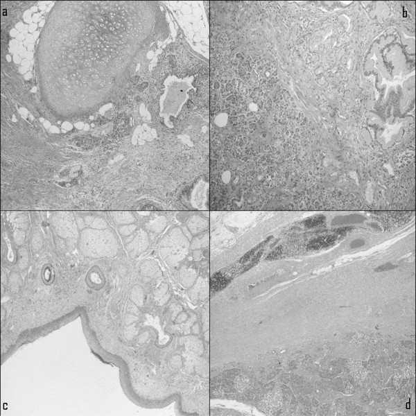
Figure 2.

Microscopic histopathological examination showing various tissue components of the mature cystic teratoma of the mediastinum. (a) Cartilaginous and adipose tissue admixed with smooth muscle fibers and lined by squamous and respiratory epithelium. (b) Pancreatic tissue. (c) Skin appendages. (d) Periphery of the lesion surrounded by residual thymic tissue with rare Hassall's corpuscles.
