Figure 3.

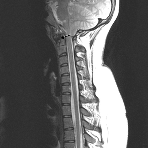
Sagittal T2-weighted MRI of the cervical spine demonstrates widening of the atlantodental interval, cervical canal stenosis without spinal cord signal changes, and pannus formation (arrow).

Sagittal T2-weighted MRI of the cervical spine demonstrates widening of the atlantodental interval, cervical canal stenosis without spinal cord signal changes, and pannus formation (arrow).