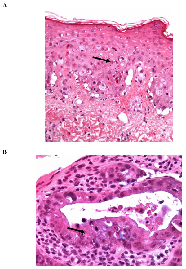
Figure 1.
Photography of skin and colon microscopic pathology. A) Skin biopsy (hematoxylin and eosin stain, ×200): an important epidermal apoptosis (black arrow) associated with basal cell hydropic degeneration can be seen in the epidermis. B) Colon: (hematoxylin and eosin stain, ×400): an increased number of apoptotic bodies combined with an increased cellular regeneration can be seen; crypt proliferative zones include many apoptotic bodies (black arrow).

