Figure 3.
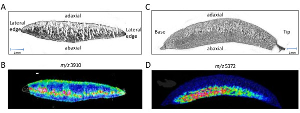
MALDI-imaging in soybean cotyledons. (A) A cross section of the cotyledon, cut across the cotyledon exposing the centre midway between the tip and the base. (B) MALDI-MSI cross section showing the peak intensity and localisation for m/z 3918. (C) Cross section of the cotyledon cut axially from tip to base. (D) MALDI-MSI cross section showing the peak intensity and localisation for m/z 5372.

